To better understand the core issues confronting the profession today, we picked the brains of some of the dental world’s leading opinion leaders and subject experts who graced the event.
By Danny Chan
The World Dental Congress, a globe-trotting event organised by the FDI World Dental Federation, offers an annual platform that nurtures ties and fosters unity among the global oral health community.
The heart of this congress, the Scientific Programme, recently unfolded over four invigorating days. With over 200 hours of insightful presentations from dental luminaries, this program served as a window into the ever-evolving landscape of dentistry. Speakers from around the world covered a diverse array of dental topics, ensuring that there was something for every attendee.
To better understand the core issues confronting the dental profession today, we had the privilege of speaking with some of the key opinion leaders and subject experts who graced the event. From periodontitis and mental health to technological advancements in endodontic diagnosis, and from environmental responsibility to the exciting world of 3D printing in oral and maxillofacial reconstruction, these presentations unveiled a tapestry of knowledge and challenges.
Through the lenses of these distinguished professionals, we delve into what lies close to their hearts and, in turn, gain insights into the predominant issues that shape the world of dentistry.
DRA Journal’s Managing Editor, Danny Chan sat down with a formidable lineup of speakers, each addressing a unique facet of the dental world:
It’s okay to ask: “Are you OK?”: Prof. Ivan Darby sheds light on the nexus between “Periodontitis and mental health,” a topic that transcends mere dental practice, reaching into the very core of human well-being. Prof. Darby advises the dental community to openly discuss mental health with patients and urges researchers to delve deeper into mechanisms like the oral and gut microbiomes.
Impact and Challenges of CBCT Imaging in Endodontics: Prof. Geoffrey Young invites us to “See around corners – the role of Cone Beam CT imaging in endodontic diagnosis and treatment planning.” The evolution of CBCT integration into routine dental practice not only streamlines our workflow but also significantly impacts patient care, unveiling a new dimension in the world of endodontics.
Search for Amalgam Replacement Long Overdue: Prof. Hien Chi Ngo extends an invitation to explore “The Minamata Convention and direct restorative options,” beckoning us to consider an alternative to dental amalgams that considers the practice environment, among other things.
Safeguarding Our Foundations: Prof. Ki-Tae Koo’s “Alveolar ridge preservation in compromised extraction sockets” emanates from a deep understanding of socket biology and their behaviour culminating in the development of alveolar ridge preservation procedures, a transformative approach that leverages the potential for enhanced healing and predictability immediately after tooth extraction.
Unveiling the Systemic Impact: In his presentation on the intriguing link between “Streptococcus mutans causing systemic diseases,” Prof. Kazuhiko Nakano highlighted the profound connection between oral health and systemic well-being, in particular, the association between oral streptococcal species and cardiovascular and cerebrovascular diseases, where Streptococcus mutans, a primary caries pathogen, plays a significant role.
Early Detection of Peri-implant Complications: Dr. Jason Pang’s workshop presentation at the FDI World Dental Congress delves into the topic of “Oral Microbiome and Peri-implantitis.” Explore Dr Pang’s proactive approach in the management of the oral microbiome and detecting complications, particularly peri-implant mucositis, at an early stage to prevent the development of implant periodontitis.
A Glimpse into Tomorrow: Prof. Richard Su unveils the intriguing world of “3D printing in oral and maxillofacial reconstruction,” leading us into the domain of future possibilities and innovation.
Pandemic Alert: Prof. Lakshman P. Samaranayake, looks back on the “Year in Review” alongside an international panel and their retrospective discussion. As one of the world’s top oral microbiologists, Prof. Samanarayake emphasises the importance of preparation for future pandemics, borrowing lessons learned during the pandemic that have transformed the dental profession.
FDI WDC Presenter: Prof. Ivan Darby (Australia)
Topic: Periodontitis and Mental Health
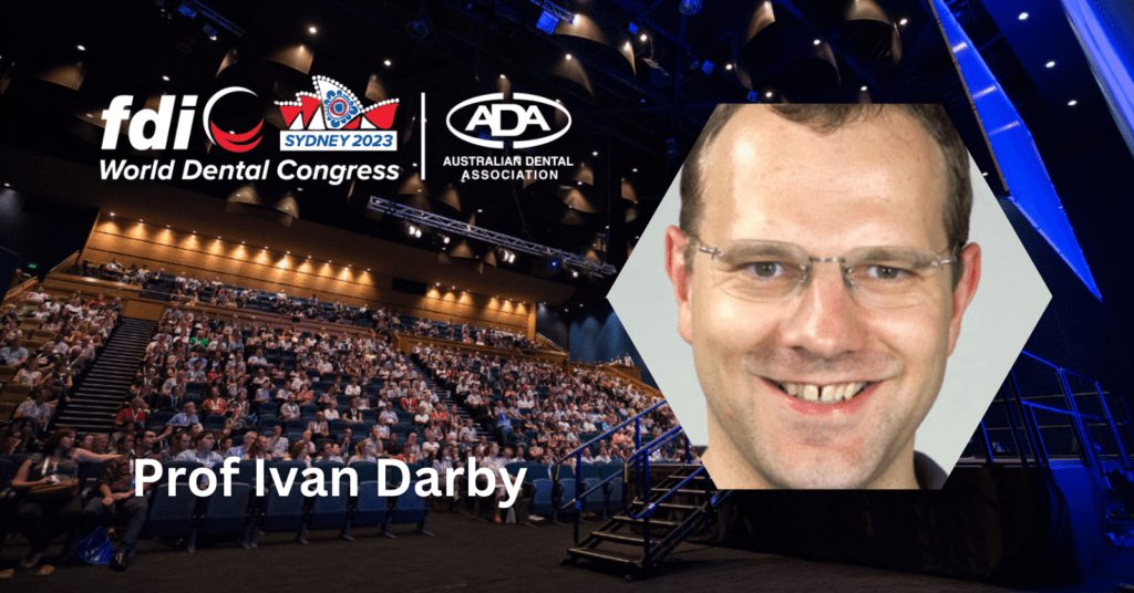
Your presentation ”Periodontitis and mental health” sheds light on the relationship between oral health and mental well-being. Why did you choose this topic?
I wanted to develop something new for this conference. This article we put together examines risk factors, and it’s an emerging field that we need to discuss. FDI gave me the opportunity to present this, and it’s close to my heart due to my family history and the many patients I’ve seen with mental health problems or on medications. We need to better manage these patients.
Do you think that oral health practitioners often overlook this issue, and is it compounded by dentists’ lack of awareness?
Yes, possibly patients need to be more aware of this, but dentists and health therapists definitely should be. Our role as health practitioners extends beyond the mouth, and we must consider the whole body. Many practitioners avoid discussing these topics, but we should address them, not just diabetes, but other co-morbidities as well.
I use deliberate language (such as pointing to a person’s weight problem) to provoke reactions and reveal what people are comfortable discussing. There are established systemic links in the literature, but we need to overcome our hesitations and talk openly about these issues, including mental health.
For the dental community, openly discuss mental health with your patients. Don’t shy away from these conversations; patients appreciate it. For researchers, delve deeper into the mechanisms, such as the oral and gut microbiomes. Explore possible avenues of treatment, like managing inflammation.
Prof. Ivan Darby
If we consider existing medical models, particularly in cases like diabetes, there are likely three key aspects to address. First, healthcare professionals should strive to understand the connections between oral health and mental health issues. Second, mental health professionals should also gain insight into these connections. Lastly, patients with mental health concerns need to be informed about the links between oral health and overall well-being.
Traditionally, general practitioners are somewhat aware of these connections, but mental health providers often prioritise the primary issues. While it’s true that for patients with severe mental health problems, concerns like periodontal disease might seem minor, it’s essential not to neglect oral health altogether. Furthermore, patients themselves often lack sufficient information about conditions like diabetes and periodontal disease, and this likely extends to mental health issues as well.
Given your extensive career, what research priorities do you see in periodontal technology, particularly concerning its impact on mental health, and how can these priorities address broader challenges?
Research priorities have evolved over the years. Mental health has been somewhat overlooked, but it’s now gaining attention. We should explore more holistic patient management as we move forward.
In the educational sphere, there should be more emphasis on these topics before students graduate.
In Melbourne, both DDS and BLS students are taught about periodontal-systemic links, especially related to smoking and diabetes. We should consider incorporating the latest research, including mental health, into the curriculum, emphasising the interrelationship between oral and overall health.
Do you believe there’s a risk of researchers being overly enthusiastic about establishing links?
No, there’s no evidence of overzealousness. Researchers are exploring a logical extension of existing knowledge in this new area. It’s a legitimate field of study with promising avenues for further research.
What advice would you offer the dental community and researchers in exploring this connection, and what does the future hold for this area of study?
For the dental community, openly discuss mental health with your patients. Don’t shy away from these conversations; patients appreciate it. For researchers, delve deeper into the mechanisms, such as the oral and gut microbiomes. Explore possible avenues of treatment, like managing inflammation. The future holds exciting opportunities to better understand and manage the relationship between oral health and mental well-being.
FDI WDC Presenter: Prof. Geoffrey Young (Australia)
Topic: Seeing around corners – the role of Cone Beam CT imaging in endodontic diagnosis and treatment planning
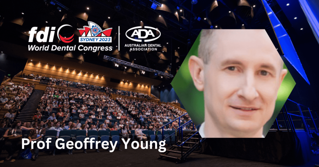
Your presentation on “Seeing around corners: the role of cone beam CT imaging in endodontic diagnosis and treatment planning” highlights the importance of advanced imaging in endodontics. What do you think are the most significant challenges in integrating CBCT technology into routine dental practice? And how do you see it impacting patient care?
Well, firstly, I must say that in terms of its impact on patient care, CBCT technology is truly exceptional. It has completely revolutionised our approach to endodontics. Until recently, we relied primarily on two-dimensional information from conventional radiographs. We did our best to work with this limited data. However, with three-dimensional imaging, we can now perceive the intricacies in three dimensions, including the buccal and palatal aspects.
This transformative technology significantly enhances our interpretation and diagnostic capabilities. Research has shown that it positively influences our treatment decisions, making us more proficient diagnosticians. It boosts our confidence in our treatment plans and even leads to changes in our treatment approach in about half of the cases. Without CBCT, we often miss crucial details. Therefore, I believe it has become an essential piece of equipment for specialist endodontic practices. As I mentioned during the lecture, one key takeaway is that in the world of endodontics, you don’t know what you don’t know until you see it in 3D. The impact of this imaging can be truly astonishing.
Regarding the barriers to integrating CBCT technology, it’s important to note that this technology has rapidly evolved. Until about 5 to 10 years ago, we were following a medical model where we had to refer patients to external radiology centres for CT scans. This disrupted our workflow significantly. We had to assess the patient, realise the limitations of 2D imaging, stop the appointment, issue a referral, and have the patient visit a radiology center. Often, the resulting images were suboptimal for endodontic diagnosis because they used large field-of-view scans with limited imaging resolution. After a waiting period of around two weeks, we would finally receive the radiology report and understand what was happening. This process was highly inefficient and cumbersome, leading us to apply it only to a select few cases.
(The medical radiology experts) are discussing radiation doses ranging from three to 50 millisieverts as being safe. In dental practice, we employ 15 to 50 microsieverts for dental CT scans, which is a thousand times lower than the proposed safe limit.
Prof. Geoffrey Young
… the evidence pertaining to the risks posed to patients by dental CT scans is somewhat outdated. … I foresee that, in the next few years, some of these restrictions might begin to be relaxed as we reevaluate the risks associated with low-dose radiation exposure.
Today, the landscape has changed. We aim to have CBCT technology integrated into our practices, which has led to a transformation in our workflow. We now acquire CBCT images at the beginning of the diagnostic process, providing us with valuable insights about the patient’s condition. This new approach allows us to be better prepared and informed, with roughly 90% of the necessary information available before we even start the treatment.
While cost and space requirements were initial barriers, these factors are gradually becoming less prohibitive. Technology has advanced significantly, making CBCT more accessible. However, one remaining concern is radiation dose. Balancing the benefits and risks of using CBCT technology is crucial. Radiation exposure is a primary point of contention, but there is a growing consensus, both in the medical and dental communities, that the doses used in low-dose CT imaging are generally considered harmless. As a result, CBCT technology is currently one of the fastest-emerging advancements in the field of endodontics.
You mentioned about a shift from relying on traditional radiography to incorporating CBCT technology in endodontics? When do you think this transition started?
It appears that one unintended consequence of the COVID-19 pandemic has been the accelerated adoption of CBCT in dental practices. Historically, there has often been a reluctance within the field of endodontics to embrace new technologies. Many endodontists were comfortable with their traditional methods and saw little need for change. Some held the belief that if they were already planning to treat a tooth, they would discover everything they needed during the procedure without the need for CBCT. This mindset was prevalent among a significant portion of endodontists.
However, the COVID-19 pandemic brought about a shift. Recommendations from organisations like the ADEA (Australian Dental Education Association) aimed to reduce the use of intraoral radiography due to the increased risk of viral transmission when patients opened their mouths during the pandemic. As a result, there was a push to use alternative imaging methods, such as CBCT, which do not require placing instruments in the patient’s mouth. This was particularly relevant for emergency cases.
As endodontists began routinely using CBCT for emergency patients based on these recommendations, they started realising the numerous benefits it offered. CBCT not only improved workflow efficiency but also provided a level of accuracy and insight that traditional radiography could not match. This positive experience led to a permanent shift in many practices towards incorporating CBCT as a standard tool.
In essence, the pandemic forced the dental community to explore new technologies, and the advantages of CBCT became evident, resulting in its increased adoption. This change has fundamentally improved the diagnostic and treatment capabilities of endodontists.
As for enhancing interdisciplinary collaboration, CBCT has proven valuable in this regard as well. It empowers endodontists to be better diagnosticians, detecting various issues such as osseous pockets, periodontal diseases, cystic lesions, and non-endodontic pathology. This enhanced diagnostic ability makes it easier for endodontists to collaborate with other dental specialists, including oral surgeons and prosthodontists, leading to improved patient outcomes. CBCT’s digital nature also simplifies the sharing of imaging data among dental professionals, further facilitating collaboration.
What emerging trends or innovations have you observed that could enhance CBCT utility in endodontics in the coming years?
I believe what we’re going to witness is the increasing prevalence of CBCT technology in specialist endodontic practice. However, a factor that has been somewhat hindering us is the consensus statement issued by the American Association of Endodontists and Oral Maxillofacial Radiologists.
While this statement dates back to 2016, it still influences our approach. Their recommendation is cautious, asserting that the risk-to-benefit ratio for routine use as a screening tool remains relatively high. Instead, they advise us to exercise individual judgment regarding its application in treatment planning. The challenge we face is that quantifying the benefits to the patient is challenging until you’ve seen the imaging because you can’t discern what you might have otherwise missed.
Over the past few years, the medical field, particularly radiology, has seen a growing chorus of voices emphasising that all radiation poses danger, adhering to a linear non-threshold model where even minimal exposure is considered risky. However, there’s a developing viewpoint that suggests there might be a threshold below which radiation is harmless. Consequently, there’s a reevaluation of the concept of applying the ALARA (As Low As Reasonably Achievable) principles. I consider this reassessment a positive development.
Regarding virtual visibility for endodontic diagnostic work, this debate is predominantly occurring within the medical sphere. They are discussing radiation doses ranging from three to 50 millisieverts as being safe. In dental practice, we employ 15 to 50 microsieverts for dental CT scans, which is a thousand times lower than the proposed safe limit.
This leads me to believe that the evidence pertaining to the risks posed to patients by dental CT scans is somewhat outdated. In response to your question, I foresee that, in the next few years, some of these restrictions might begin to be relaxed as we reevaluate the risks associated with low-dose radiation exposure.”
FDI WDC Presenter: Prof. Hien Chi Ngo (Australia)
Topic: The Minamata Convention and direct restorative options
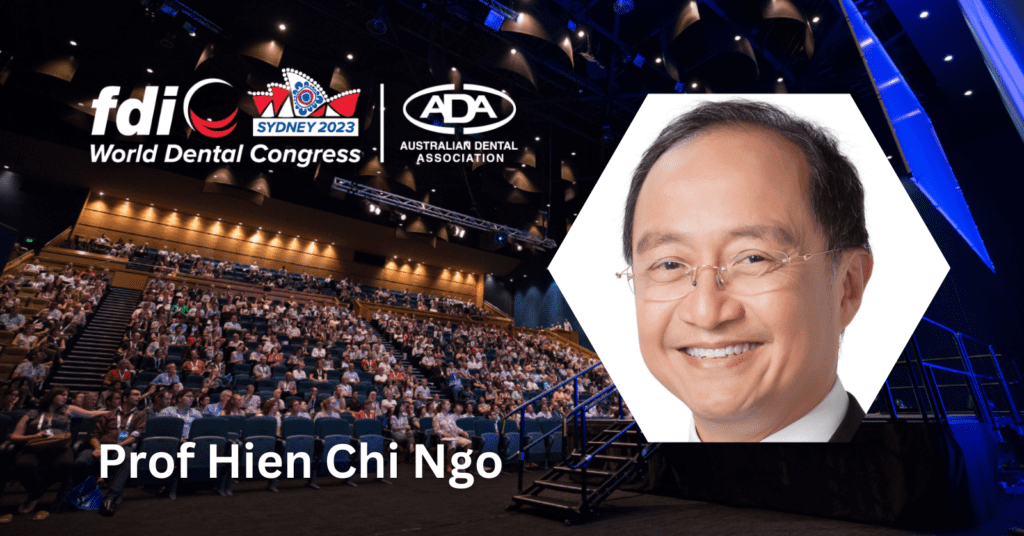
Can you share with us why you chose to focus on “The Minamata Convention and direct restorative options” in your presentation? What led you to this particular topic?
I selected this topic because the European Union is considering to ban amalgam by 2025, which is just two years away. The key issue here is that we do not have an amalgam replacement ready
It depends on the clinical environment. If you can control moisture and your clinical working conditions, alternatives like composite and glass ionomer seem feasible. However, in rural areas or developing countries where infrastructure is lacking, a material that is easy to handle and cost-effective becomes crucial. I believe we haven’t yet found that material. Therefore, we need to engage in discussions and provide guidance to manufacturers on the properties and characteristics such a material should possess before it can be considered a true alternative to amalgam.
My lecture aimed to enhance people’s understanding of glass ionomer cements, as they are easier to handle and more technique-tolerant. This could potentially form the basis for developing a material suitable for amalgam replacement in situations where the clinical working environment is less than ideal.
Can you provide more details about your earlier statement regarding GICs and their potential role?
Currently, we know that glass ionomer cements perform well when used in the right cases and handled correctly. Being water based, GIC is less technique sensitive than composite resin, when it comes to moisture control during placement. This is an important advantage over composite resin.
Composite resin can work effectively, but it has specific requirements that must be met for optimal performance. In contrast, glass ionomer cement is technique-tolerant and easy to work with. We also have extensive long-term clinical experience with GIC, demonstrating its reliability. Considering these factors, I would say that GIC has more potential as an amalgam replacement than composite resin, especially when it comes to ease of placement and low cost.
It’s crucial for our profession to come together and define the necessary attributes of a material before it can be considered a true alternative to amalgam. This will provide a clear framework for manufacturers to work within and design such materials effectively.
In the context of choosing the right amalgam replacement that is suitable for developing countries and rural areas, could you elaborate on the factors that limit the consideration of some other materials and explain why GIC possesses some of these favourable attributes?
Firstly, the cost of using composite resin is higher than that of glass ionomer cement (GIC) for dental restorations. Cost becomes particularly significant when we think about developing countries. Secondly, when it comes to technique sensitivity, amalgam is quite forgiving, and GIC is also fairly tolerant of variations in technique. In contrast, composite resin is the least forgiving in terms of technique sensitivity. Lastly, consider the skill set required for dentists to work effectively with these materials. Training dentists to perform composite resin restorations well often requires more effort and expertise than training them to work with amalgam or GIC.
EDITOR’S PAGE | ADVISORY BOARD | NEWS | PRODUCTS | FEATURE ARTICLE | CLINICAL | PROFILE | EXHIBITIONS & CONFERENCES | PRODUCT TIPS | DENTAL BUSINESS
GICs not only offer a suitable amalgam replacement in developing countries. For example, if you work in a rural area in Austria or Australia. Even there, you might face situations where you have to work single-handedly. In those scenarios, you have to consider materials that are more technique-tolerant, especially when you don’t have additional assistance. Imagine a day when your air-water syringe malfunctions and starts spitting out water, and you call the technician, but they can’t arrive for 48 hours because you are far from the city. How can you function for the next few days if you are using a material that requires a dry environment? In such situations, glass ionomer cement (GIC) may be the only feasible solution available, as it can perform adequately even in less-than-ideal conditions. We need to consider not only clinical selection but also our working environment because conditions can change, and we must have the right tools available to adapt to them.
FDI WDC Presenter: Prof. Ki-Tae Koo (South Korea)
Topic: Alveolar ridge preservation in compromised extraction sockets
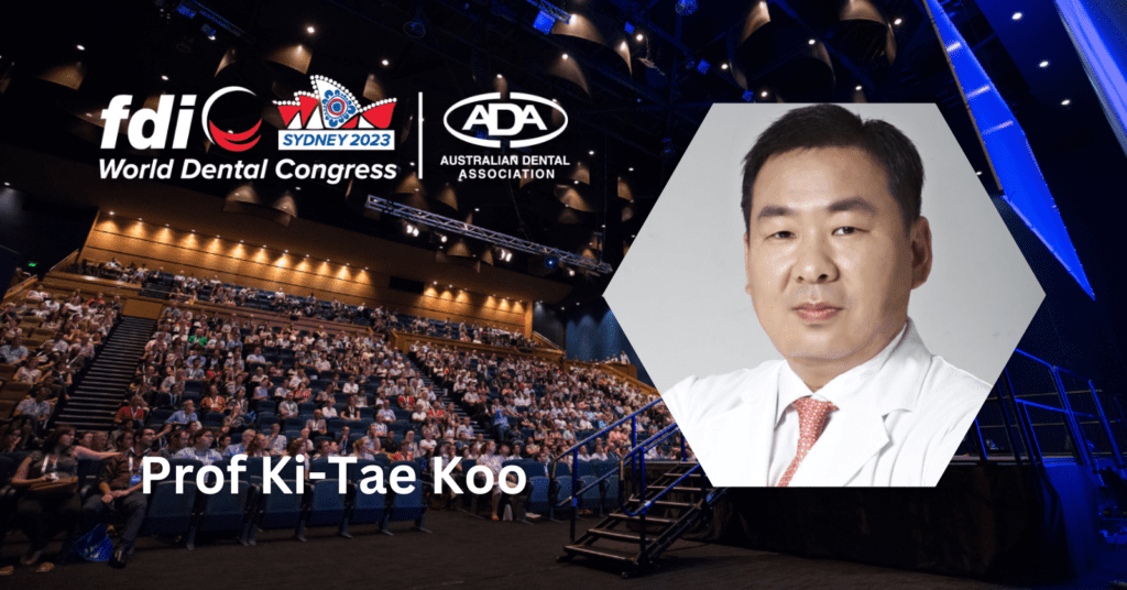
Why did you choose this topic for the FDI Congress, and why does it hold particular significance?
I began to develop an interest in socket biology during my training years, which dates back to my time at Temple University in the USA. My mentor, Dr. Will Fitzpatrick, was a master in the field of wound healing, specifically periodontal wound healing. I believe that’s where it all started for me. When I returned and began my career in teaching and research at the Seoul National University, I started to delve into the healing of extraction sockets. We began investigating what happens after tooth extraction, focusing on dimensional changes and socket healing. Some sockets exhibited excellent healing, while others did not. This piqued our curiosity, leading us to explore erratically healing sockets – those that did not heal on their own despite various efforts.
Our research journey encompassed preclinical and clinical studies on erratically healing sockets, ultimately leading to ridge preservation procedures. When you perform ridge preservation immediately after tooth extraction, you capitalise on greater healing potential and predictable outcomes. It’s important to note that I wouldn’t recommend ridge preservation in sockets that would heal naturally. That’s a definite no-no. However, for sockets that show signs of erratically healing, ridge preservation procedures can save time for both the patient and the practitioner while maintaining a strong patient-dentist relationship.
I don’t believe we have a clear answer to the question of what can replace amalgam. It depends on the clinical environment. If you can control moisture and your clinical working conditions, alternatives like composite and glass ionomer seem feasible.
Prof. Hien Chi Ngo
However, in rural areas or developing countries where infrastructure is lacking, a material that is easy to handle and cost-effective becomes crucial. I believe we haven’t yet found that material.
Our journey began with socket biology, delving into clinical procedures for erratically healing sockets, and now we’re focusing on infected and compromised sockets. We aim to understand the characteristics of compromised sockets, how they heal, and how we can overcome the associated challenges. My response to this challenge is a resounding yes – predictability is high for single sockets. Based on our success, we’re gradually expanding our approach to encompass multiple extractions and pockets.
In the long run, this approach may potentially reshape the paradigm of guided bone regeneration. Typically, after extraction, we allow sockets to heal without intervention, resulting in the loss of soft and hard tissues, along with dimensions such as volume and width. Our goal is to prevent this loss by preserving what’s present at the time of extraction. In summary, this approach simplifies the process for everyone involved, provided we can effectively control infection.
What are some of the most promising advancements or techniques in alveolar ridge preservation?
I believe that biomaterials have made significant advancements, resulting in improved predictability. These advancements have led to better outcomes, such as enhanced new bone formation, reduced risk of infection and reinfection, and favourable long-term results. Specifically, there is less shrinkage, bone loss, and dimensional changes over time. These materials can maintain their integrity, contours, height, and other aspects over the long term. In summary, they have become quite predictable.
When you perform ridge preservation immediately after tooth extraction, you capitalise on greater healing potential and predictable outcomes. It’s important to note that I wouldn’t recommend ridge preservation in sockets that would heal naturally. That’s a definite no-no. However, for sockets that show signs of erratically healing, ridge preservation procedures can save time for both the patient and the practitioner while maintaining a strong patient-dentist relationship.
Prof. Ki-Tae Koo
With regards to your affiliations with prestigious institutions in South Korea and the United States, how has your international exposure and collaborative work influenced your perspective on topics like bone regeneration and implantology? And how does this contribute to the global conversation on these subjects?
My international exposure has mainly come through involvement with significant organisations like the Osteology Foundation, where I serve as an Expert Council member and a fellow. However, it remains somewhat limited due to our geographical location in Asia. While our scientific contributions, evidence-based research, and clinical work are strong, there are still limitations, possibly due to language barriers, especially in English-dominated discussions. Nevertheless, I do see improvements over time.
Opportunities for Asians in the field are expanding, and I’ve been honoured to participate in events like FDI. For instance, I had the privilege of sharing the stage with renowned periodontal experts such as Prof. Maurizio Tonetti and Prof. Mariano Sanz, which was a significant honour. The trend is toward more inclusivity, with growing opportunities for Asians to participate and contribute positively.
As scientists in Asia, our work and research are contributing to the advancement of technology and science in the region. The environment is gradually improving, offering more opportunities. Exposure on international platforms is essential because it helps gain recognition for our protocols, concepts, and developments. In essence, this exposure is quite beneficial, and I believe that as time goes on, it will continue to enhance our contributions to the field.
FDI WDC Presenter: Prof. Richard Su (Hong Kong)
Topic: 3D printing in oral and maxillofacial reconstruction
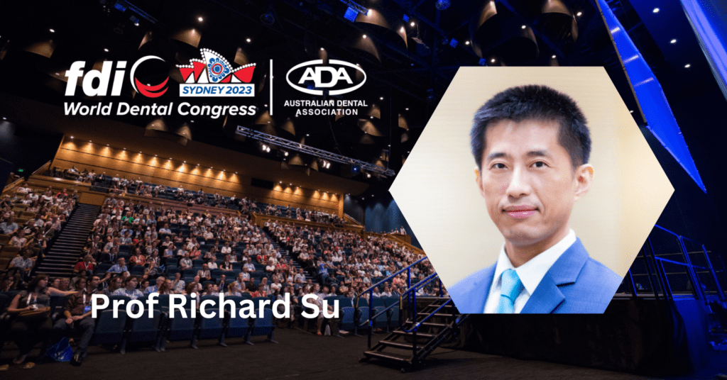
My question pertains to your extensive background in research and actual surgery involving 3D-assisted planning, as well as the printing of surgical guides and surgical prostheses. Could you please tell us why you chose this specific topic for your presentation today at the FDI?
Computer-assisted 3D printing is one of the cutting-edge technologies that has recently gained widespread use in medicine and dentistry. In my clinical practice, we extensively utilise this technology, and I consider it a game-changer. It represents a paradigm shift from traditional freehand surgery to computer-assisted surgery, where every aspect of the procedure is meticulously planned using computer software before the actual surgery takes place. This approach not only saves valuable operating time but, more importantly, significantly enhances the precision of surgical procedures.
In the past, surgery often relied heavily on the experience and skill of the surgeon, which could lead to a considerable amount of trial and error during the surgery itself. Adjustments and modifications were made on the fly to achieve the desired outcome. This method was particularly challenging for young and less-experienced surgeons due to the steep learning curve associated with it.
However, with the integration of new technology, everything is meticulously planned and simulated on the computer beforehand. Thanks to 3D printing technology, surgeons can execute surgical procedures more accurately during surgery itself. This transformative approach not only benefits young and less-experienced surgeons by helping them achieve optimal surgical outcomes, but it also changes the landscape of clinical teaching and research in the field of surgery.
In essence, the adoption of this technology has ushered in a new era, impacting every facet of surgical practice. That’s why I chose this topic for my presentation at the FDI Congress.
Open-mindedness is crucial in embracing new perspectives and ideas from different disciplines. In today’s modern medicine and dentistry, every field is highly specialised, and it’s impossible for one person to know everything. Humility and cooperation are key. Only by fostering a spirit of multidisciplinary collaboration can we advance science and improve patient care. We must welcome diverse opinions and ideas, as they contribute to our collective progress in this field.
Prof. Richard Su
You’re on the cutting edge of technology, and essentially, your work is future-oriented. What are the challenges of being in that position? Because being on the forefront means you have very few precedents to fall back on. So do you find that challenging as you move forward?
When your research is always evolving, the need to adapt and innovate is constant. Yes, it is challenging. However, it also brings inspiration, and as you overcome these challenges, you achieve progress. That’s one of the joys and the fulfilling aspects of being in academia and conducting personal research in clinical settings. With each challenge, you have the opportunity to innovate and ultimately improve patient outcomes. The ultimate goal is to enhance treatment outcomes for our patients, and we achieve this by embracing new knowledge and drawing insights from various disciplines.
We engage in interdisciplinary collaboration, and yes, it can be challenging because different fields may have their unique languages and approaches. However, we all work toward a common goal. To navigate potential miscommunications or problems arising from this novelty, we need to be open-minded.
Open-mindedness is crucial in embracing new perspectives and ideas from different disciplines. In today’s modern medicine and dentistry, every field is highly specialised, and it’s impossible for one person to know everything. Humility and cooperation are key. Only by fostering a spirit of multidisciplinary collaboration can we advance science and improve patient care. We must welcome diverse opinions and ideas, as they contribute to our collective progress in this field.
What do you think is the future of computer-assisted surgery, 3D printing, and 3D planning in the realm of dental education and training?
Well, dental education is already evolving due to these technologies. The younger generation is growing up with computers, and they adapt to technology faster than previous generations. With computer-assisted surgery, achieving favourable surgical outcomes becomes easier for the younger generation, which shortens the learning curve compared to the older generation. In the future, computer systems may incorporate more artificial intelligence (AI) components, further enhancing surgical outcomes and possibly leading to more intelligent surgeries.
With AI, virtual surgical planning may become more efficient and autonomous. This means that AI could take over some of the tasks that humans currently perform in virtual surgical planning. This represents a significant advancement in technology.
You advocate for bringing all this technology in-house rather than using commercial services. What are your main reasons for this approach?
One of the primary reasons is that we are an educational institution. We aim to equip our students and young residents with hands-on experience in using these technologies. The younger generation, particularly Chinese residents, is very tech-savvy, and we believe that providing in-house access to these technologies benefits their education.
This approach differs from that of private practitioners who might prefer outsourcing to save time and resources. However, our focus is on educating the next generation of dentists and surgeons, and that’s why we emphasise the importance of learning how to use these technologies effectively.
FDI WDC Presenter: Prof. Kazuhiko Nakano (Japan)
Topic: Streptococcus mutans causes systemic diseases
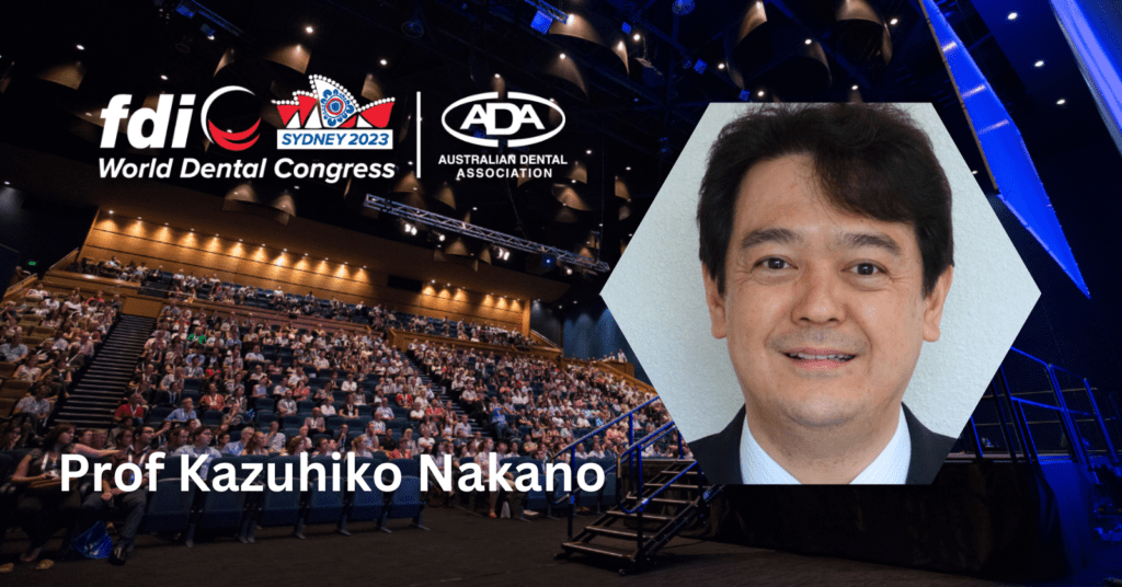
In your presentation on “Streptococcus mutans causing systemic diseases,” you highlighted the connection between oral health and systemic health. Can you elaborate on the most pressing systemic health issues that dentistry needs to address in relation to this research?
I am sure that there should be much more dissemination of information regarding the association between oral streptococcal species and cardiovascular and cerebrovascular diseases. It should be known that Streptococcus mutans, a major caries pathogen, is one of them. Reducing the bacteria in the oral cavity is crucial, especially for people with underlying heart disease. Additionally, elderly individuals with decreased immunity should also take precautions. Endothelial dysfunction can occur in people without heart disease due to various factors. Dentists should be prepared to treat such patients to maintain their oral health.
Streptococcus mutans is a well-known contributor to dental caries. How do you think the dental profession should adapt its preventive and treatment strategies in light of the emerging evidence regarding its systemic impact?
S. mutans is present in the oral cavity of most people, with varying amounts. Recent discoveries have shown that S. mutans with a collagen-binding protein called Cnm on their cell surface can exacerbate cardiovascular and cerebrovascular diseases. Currently, there is no straightforward way to identify individuals carrying these high-risk bacteria. Therefore, it is important to reduce the amount of S. mutans overall. For individuals with dental caries, treatment of the carious lesion can help reduce bacterial levels. Professional dental cleaning is another way to achieve this reduction. Furthermore, providing guidance on personal oral care at home is crucial.
Recent discoveries have shown that S. mutans with a collagen-binding protein called Cnm on their cell surface can exacerbate cardiovascular and cerebrovascular diseases. Currently, there is no straightforward way to identify individuals carrying these high-risk bacteria. Therefore, it is important to reduce the amount of S. mutans overall.
Prof. Kazuhiko Nakano
In your opinion, what collaboration opportunities exist between the fields of dentistry and other healthcare disciplines to address the systemic health issues associated with Streptococcus mutans effectively?
Data has linked Cnm-positive S. mutans to infective endocarditis, cerebral hemorrhage, vascular dementia, as well as non-alcoholic steatohepatitis, inflammatory bowel disease, and IgA nephropathy. Research on these diseases varies in development stages, but there is evidence of an association in both basic and clinical research.
It is crucial to collaborate with medical specialists in these diseases. We are also researching whether reducing the amount of Cnm-positive S. mutans in the oral cavity can improve these associated diseases. We hope to publish findings on this within the next year or two. Subsequently, we can apply the new method to a larger number of patients.
How can dental education and training be adapted to ensure that future generations of dentists are well-equipped to recognise and manage the systemic implications of oral health conditions?
The understanding of the impact of oral health on the entire body is relatively recent. It hasn’t been extensively integrated into dental education for students thus far. While much has been learned recently, there is still much to interpret.
Currently, we present research results and leave interpretation to individual students. Once interpretations are established based on different evidence, we can incorporate this into textbook education. However, it may take some time before this happens. Nevertheless, I hope to encourage people to seek various sources of information, keeping in mind that oral health affects the entire body.
FDI WDC Presenter: Dr. Jason Pang (Australia)
Topic: Oral microbiome and Peri-implantitis
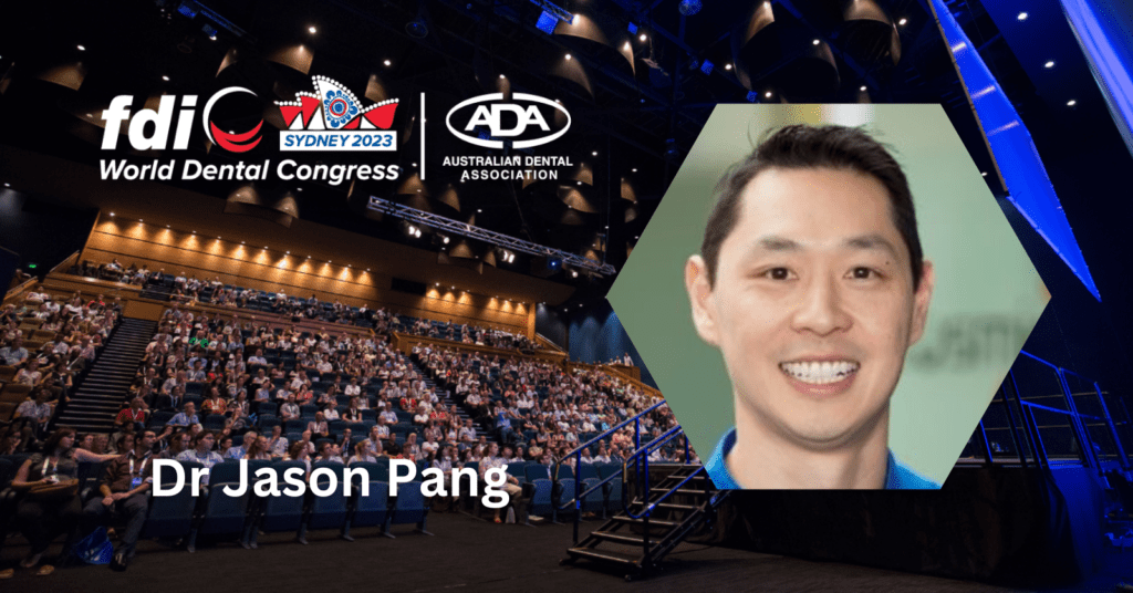
Why did you choose this topic for your workshop presentation?
The topic is about managing the oral microbiome and early detection of complications, specifically peri-implant mucositis, to prevent implant periodontitis. Instead of waiting for symptoms like pocketing, bleeding, and bone loss, we can use a phase contrast microscope to identify species likely to cause complications. For instance, if we observe a significant presence of spirochetes, such as Treponema DeNicola, along with white blood cells, we can identify individuals at risk of complications. This allows us to detect and address issues months, or even years, before they progress to severe bone loss, ultimately preventing major problems.
Peri-implant complications are a global concern for practitioners, and currently, there’s no definitive treatment for periodontitis. However, the best approach we have is managing implant mucositis early to prevent implantitis. Therefore, early detection and treatment are crucial to addressing these issues before they escalate.
Peri-implant complications are a global concern for practitioners, and currently, there’s no definitive treatment for periodontitis. However, the best approach we have is managing implant mucositis early to prevent implantitis. Therefore, early detection and treatment are crucial to addressing these issues before they escalate.
Dr. Jason Pang
You are also a strong advocate and trainer in the field of laser dentistry. Are you satisfied with the laser-related presentations, or do you believe there’s room for improvement in this regard?
I believe FDI is relatively new to laser dentistry, and the introduction of a few laser-related lectures is a positive step. However, there is certainly room for more in-depth discussions on lasers in dentistry. Laser applications extend beyond merely cutting gum tissue. Presentations like George Romanos’ discussion on lasers and peri-implantitis, and A/Prof. Kinga Grzech Lesniak’s insights on periodontitis are all valuable topics. It’s essential for dentists to understand how lasers can be integrated into daily practice, going beyond their conventional uses.
FDI WDC Panelist: Prof. Lakshman P. Samaranayake (Hong Kong)
Topic: The Year in Review
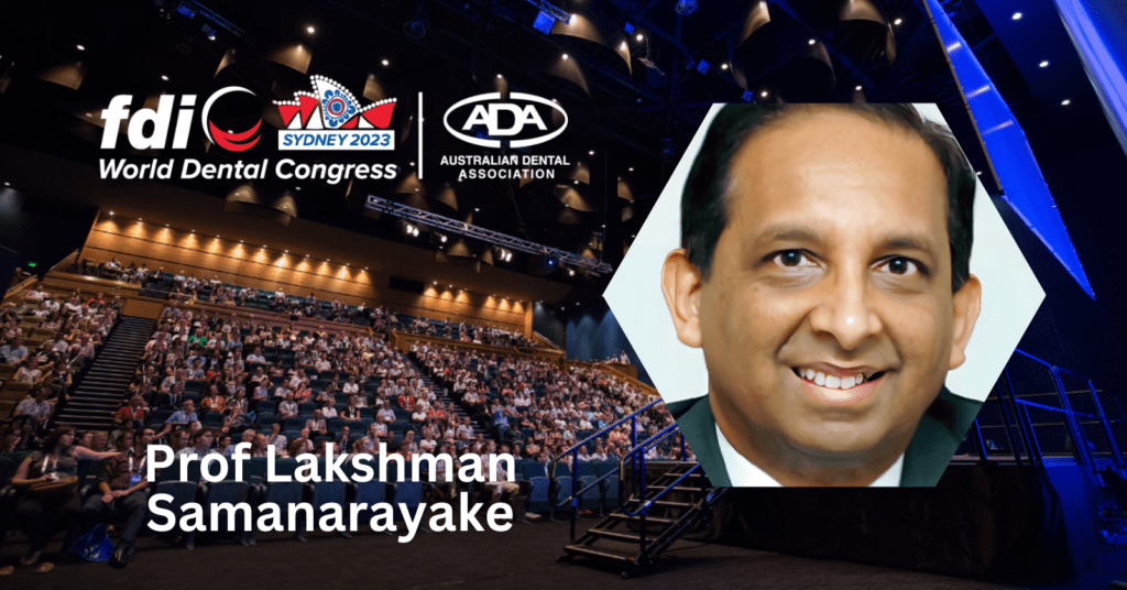
In your panel discussion, what were the most prominent trends and developments in the field of dentistry that emerged in 2023?
During the panel discussion, we had participants from different parts of the world, including the UK, US, Australia, and myself from Hong Kong. Each of us shared our unique perspectives on how dentists and the public were affected in our respective regions, as well as how people were coping with the current situation and the prospects for the future.
The chairman posed various questions regarding these aspects. From what I gathered, there were no significant surprises, as the dental profession worldwide was severely affected by the ongoing pandemic. This impact was felt through the closure of medical clinics in various parts of the world, leading to a significant backlog of untreated illnesses. Unfortunately, this neglect extended to diseases such as oral cancers because patients were reluctant to visit dental clinics out of fear of contracting COVID-19.
One noteworthy aspect discussed was the use of digital platforms like Zoom for information dissemination. Prominent speakers and opinion leaders from different countries, including myself and Prof Purnima Kumar from the USA, participated in Zoom meetings with thousands of attendees. This created a new avenue for sharing information about infection control and other dental-related topics.
Additionally, we shared personal experiences of how COVID-19 affected us individually, where we were during the pandemic, and how we learned about it. These personal stories highlighted the challenges faced by families, including the inability to see loved ones, especially grandchildren. Surprisingly, few panelists discussed financial losses, possibly because most of them were affiliated with universities and continued to receive their salaries during this period.
In summary, the panel discussion provided valuable insights into the global impact of the pandemic on the dental profession and showcased the use of digital platforms for information exchange.
There should be a direct line of communication with organisations like the WHO or other global health bodies, where representatives from FDI and other organisations can convey these directives. A unified response will be more authoritative and less confusing. I believe we should find a way to establish a coordinated response during pandemics, involving regional, local, and international dental organisations, as well as the WHO.
Prof. Lakshman P. Samaranayake
From your perspective as one of the top oral microbiologists in the world, and given your significant role in the field of dentistry, what are your personal thoughts on the outlook for dentistry this year? And what message would you like to convey to your colleagues worldwide as they navigate the challenges and opportunities ahead?
As a clinical microbiologist, I see both positive and negative aspects in our profession in the need to be well-prepared for future pandemics. Firstly, we have made significant progress in terms of infection control. However, there are still areas where improvement is needed. Secondly, vaccination is crucial. Dentists and the entire dental team should strictly adhere to vaccination protocols, not only for COVID-19 but also for other infectious diseases. It’s essential to keep accurate vaccination records. In some countries like the UK, failure to maintain these records can lead to legal issues through inspections and other professional bodies.
Thirdly, the dental profession has become more conscious of the importance of proper infection control. They faced financial hardships during the pandemic due to practice closures, which has served as a valuable lesson. Dentists are now taking infection control procedures more seriously.
Lastly, educating the public about dentistry’s safety is vital. Visiting a dentist is no more dangerous than visiting a doctor. Neglecting oral diseases can lead to severe consequences, including death in cases of cancer. It also results in increased expenses as the disease progresses. Overall, dentists have learned important lessons from the pandemic, making them more aware and conscious of infection control compared to 20 or 30 years ago.
My message is that we must be extremely cautious and take all necessary precautions, including using personal protective equipment, getting vaccinated, and implementing robust infection control procedures. These three areas are critical for maintaining high standards in dental practice.
My final question stems from the fact that you have been a long-standing supporter of FDI. Not only are you the sitting chief editor of the International Dental Journal (IDJ) – the flagship publication of FDI – you’ve also held various office bearer positions at many levels throughout the years. FDI is one of the few dental organisations that unites the world through national representation. From your perspective, what are your thoughts about FDI and events like the FDI World Dental Congress that convene and address the challenges facing the dental profession in various dental communities worldwide, and whether it serves as a valuable platform for collaboration and knowledge sharing?
As the editor-in-chief of International Dental Journal (IDJ), I often come across manuscripts, like one from from a developing nation, where they investigated how the COVID-19 pandemic had impacted the dental industry. Interestingly, there have been a notable number of dentist deaths in that country – around 20, I believe. While it’s challenging to attribute these deaths directly to dental practice, it appears relatively high in that region. They attribute this partly to controversies regarding infection control recommendations between the Health Department and the government, as well as conflicts with the professional dental association. People were left uncertain about what to do, and the government’s support was lacking.
My point here is that we must unify our efforts. Authoritative bodies like FDI, with their wealth of expertise, can disseminate guidelines. There should be a direct line of communication with organisations like the WHO or other global health bodies, where representatives from FDI and other organisations can convey these directives. A unified response will be more authoritative and less confusing. I believe we should find a way to establish a coordinated response during pandemics, involving regional, local, and international dental organisations, as well as the WHO.
FDI’s already established extensive network of National Dental Associations (NDAs) could be used as an emergency portal that could be linked to promulgations of not only the FDI but also international bodies such as the WHO. That’s my personal view. FDI has a vast network of NDAs, and I believe it should play a more active role, especially in light of the lessons learned during the COVID-19 pandemic.
The information and viewpoints presented in the above news piece or article do not necessarily reflect the official stance or policy of Dental Resource Asia or the DRA Journal. While we strive to ensure the accuracy of our content, Dental Resource Asia (DRA) or DRA Journal cannot guarantee the constant correctness, comprehensiveness, or timeliness of all the information contained within this website or journal.
Please be aware that all product details, product specifications, and data on this website or journal may be modified without prior notice in order to enhance reliability, functionality, design, or for other reasons.
The content contributed by our bloggers or authors represents their personal opinions and is not intended to defame or discredit any religion, ethnic group, club, organisation, company, individual, or any entity or individual.

