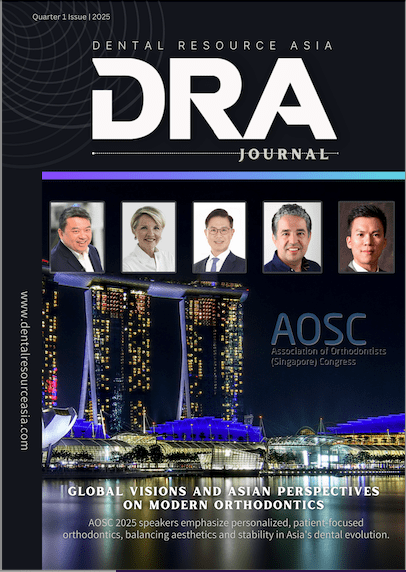
Unlock the secrets to accurate scanning as Dr. Baresel provides valuable insights and tips for dentists looking to excel in the world of intraoral scanning.
By Danny Chan
Digital technologies stand as the driving force behind transformative advancements In the dynamic world of modern dentistry. Dr. Ingo Baresel is a fervent advocate of digital dentistry and distinguished figure in this field.
The founder and president of the German Association of Intra-oral Scanning is a well-regarded opinion leader on intraoral scanning and one of the pioneering users of the iTero system from Align Technologies. Dr. Baresel’s unique position as an expert in digital dentistry and intraoral scanners, makes him an excellent candidate to share tips on the best practices of implementing intraoral scanners in your practice. Drawing from his rich experience with scanners like iTero, Trios, Straumann Virtuo Vivo, Carestream, Medit, Dental Wings, and Sirona, Dr. Baresel serves as a seasoned authority on the subject.

Dental Resource Asia: Please share your journey in the field of digital dentistry and intraoral scanning, from your early years to your current role as the founder and president of the German Association of Intraoral Scanning.
Dr Ingo Baresel: Okay, so I started my intraoral scanning career in 2012 when I purchased my first iTero scanner, the first to scan without powder. Before that, I always explored tech systems but was never satisfied due to accuracy issues, especially as I didn’t want to work with powder.
When I heard about the iTero scanner from a colleague, I made a quick decision, ordered it, and began using it in 2012. Over the years, we progressively transitioned to digital workflows, starting with small restorative work and expanding to include implants and other procedures. As new scanners entered the market, we incorporated them into our practice.
In 2014, recognising the lack of focus on intraoral scanning in existing discussions about digital workflows, we founded the German Association of Intraoral Scanning to address this gap. Subsequently, companies approached me to test and provide feedback on their scanners, leading me to gain extensive experience with almost every scanner available.
DRA: Can you tell us more about what the Association does and why you formed it in 2014?
IB: We established the association because existing dental associations primarily focused on digital lab workflows and not on the initial step of scanning. Our goal was to fill this gap and advocate for intraoral scanning. We now have members, host annual meetings, conduct workshops, and collaborate in study groups to advance digital dentistry. At the moment, we have around 150 German dentists as members.
DRA: You emphasise the importance of technical communication in dentistry. Can you elaborate on what this means in the context of modern practice with digital systems?
IB: Technical communication, especially with the lab, is crucial when initially setting up digital workflows. However, once established, working with the lab becomes more manageable. Digital systems offer additional communication options, such as sending real pictures of preparations and teeth, enhancing the collaboration between dentists and labs for improved diagnostics and treatment planning.
To begin with, transitioning to digital processes requires the initial setup of new workflows and effective communication with the dental laboratory. Once these foundational steps are in place, the advantages become apparent. Digital workflows eliminate dependence on factors like humidity and room temperature during processes such as alginate mixing.
The initial focus should be on establishing these workflows and engaging in communication with the dental lab. Once this foundation is laid, collaboration with the lab opens up numerous possibilities for conveying information effectively. For instance, when scanning a patient, both colored and monochrome scans offer clarity in observing margin lines. Specific scanners, such as those from Itero and Exocad, provide detailed images of preparations and the patient’s teeth.
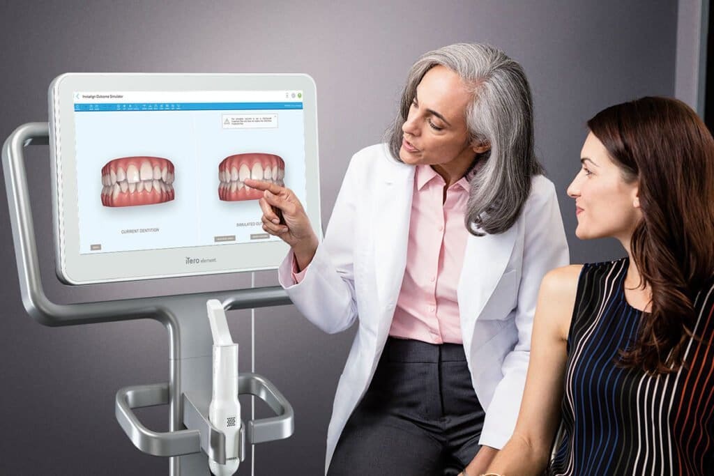
Collaboration with the lab also allows for quality checks. For extensive cases like full-mouth scans, the lab can review the scans, and any necessary adjustments can be made through conventional methods. Effective communication with the lab, particularly in intricate cases like implant workflows, enhances overall quality. In the case of implant scans, digital scanning facilitates the capture of the emergence profile, a task challenging with conventional impressions. Intraoral scanning and digital workflows, therefore, contribute to elevating the quality of dentistry, providing me with the tools to be a more proficient dentist.
The learning curve is considerably short, but dentists should commence with small cases, avoiding complex situations like implant cases initially. Starting with single premolars or similar cases is advisable. Learning to scan swiftly is paramount, and practitioners must adapt to the process.
Dr. Ingo Baresel
DRA: With approximately 10 years of experience and having worked with various scanners, what insights can you offer into intraoral scanning technology, and what might dentists overlook?
IB: I have found that the majority of dentists refrain from using scanners due to perceived issues like accuracy, cost, and complexity. The crucial message here is to debunk these misconceptions. Numerous studies, numbering in the thousands, unequivocally demonstrate the accuracy of scanning when performed correctly. Similar to conventional impressions, accuracy depends on proper technique; both methods are accurate when executed correctly.
The learning curve is considerably short, but dentists should commence with small cases, avoiding complex situations like implant cases initially. Starting with single premolars or similar cases is advisable. Learning to scan swiftly is paramount, and practitioners must adapt to the process.
During scanning, it’s essential to focus solely on the screen and not on the patient. This approach ensures independence between the movement of the wand and the visual attention on the patient. Scanning at a brisk pace is vital, especially with the latest fast scanners, as slow scanning diminishes rather than enhances quality.
DRA: Why is it recommended not to look at the patient while scanning?
IB: Looking at the patient can lead to slower scanning, and you may miss whether the scanner is still acquiring data. To ensure efficient and accurate scanning, it’s necessary to focus solely on the screen and move the wand independently.
DRA: What are your thoughts working with the iTero?
IB: One of the primary reasons I prefer working with iTero is its scanning strategy. Unlike other scanners where you need to scan all prepared teeth together, following the occlusal-lingual-buccal sequence, iTero stands out. It allows me to scan each prepared tooth individually, removing the need to handle all teeth at once, eliminating difficulties related to gingiva collection, bleeding, and labor. With iTero, I can scan one prep tooth at a time, performing a full arch scan later, seamlessly integrating high-resolution scans into the overall image. This significantly streamlines my workflow, sparing me the need for time-consuming scan repairs.
Another key feature I appreciate in iTero is its near-infrared imaging system, which aids in detecting caries lesions during each scan. This post-scan analysis enables me to identify and address any issues with caries promptly. Through our study, we found that near-infrared imaging is much more precise in identifying early caries compared to conventional methods. This early detection allows for prompt intervention and treatment using modern techniques such as proximal sealing or infiltration methods. This capability enhances my preference for working with this technology, as it facilitates early detection and contemporary treatment approaches.
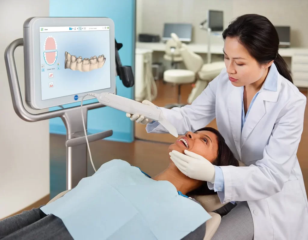
These are just two of the several reasons why I favour iTero. It’s important to note that other scanners aren’t inherently bad, and I don’t disparage them. However, for my specific workflows and to minimize stress in my professional life, the user-friendly scanning process and advanced imaging capabilities of iTero make it my scanner of choice.
DRA: How is iTero, in conjunction with near-infrared images, utilized over time for comparison?
IB: Currently, we have the functionality to compare scans over time, primarily for assessing tooth movement, abrasion, and changes in gingival areas. The scanner overlays two scans, detecting alterations and presenting them visually to the patient. Measurements can also be taken using the near-infrared images.
However, as of now, there isn’t a time lapse function specifically for near-infrared imaging. While you can compare two scans manually, future developments may introduce a time lapse feature for near-infrared imaging, allowing for a more comprehensive comparison of the sizes of carious lesions over time.
DRA: How do intraoral scanners promote better dentist-patient communication?
IB: it’s quite simple – these tools enable the early detection of issues compared to traditional bitewings, a fact supported by studies. Early identification provides an opportunity to engage with the patient proactively. By visually presenting findings, such as white spots, patients better comprehend the necessity for treatment – seeing is believing. This proactive communication allows patients to understand the reasons behind treatment recommendations.
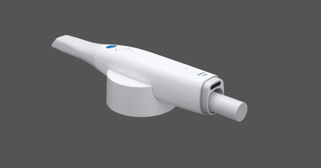
In the case of early caries, not visible on bitewings, the practitioner has options: waiting and monitoring or offering preventive treatments like proximal sealing or infiltration. This decision-making process involves the patient, presenting a choice between monitoring and proactive treatment to prevent future issues. This diagnostic approach proves highly beneficial for me, aiding in effective communication with patients about their needs and the reasons behind proposed treatments.
DRA: For those considering adopting digital technology, what tips or pitfalls should they be aware of?
IB: The first piece of advice is not to attempt everything simultaneously. There’s no need to invest in a scanner, printer, or milling machine all at once. Most labs, especially those working with systems like CEREC or E4D, are already digitally equipped.
Therefore, the initial step towards digital adoption is to digitize impressions, necessitating the acquisition of an intraoral scanner. The market offers numerous options, each with distinct features, so selecting the right one is crucial. After choosing a scanner, the next step is gradual integration into your workflow.
Starting with simpler cases before progressing to more complex ones is advisable. As confidence in the technology grows, you can gradually expand your usage, eventually considering additional investments such as a chairside milling machine or a 3D printer for specific applications like nightguards. The key takeaway is to start by digitizing impressions and then progressively explore further digital applications.
Start gradually, beginning with intraoral scanning to digitize impressions. Seek advice from experienced individuals who have used various systems. Consider accuracy and ease of handling rather than solely focusing on price. Progress step by step, starting with simpler cases before expanding to more complex procedures.
DRA: How would you recommend someone starting the process of purchasing an intraoral scanner?
IB: This is a challenging question as there are few individuals globally who have the opportunity, like myself, to evaluate and understand the benefits and drawbacks of every scanner. My advice is to seek out someone with extensive experience across various systems and heed their advice. However, it’s important to note that individual experiences with different scanners vary, and recommendations often align with personal preferences. While it may not be easy, try reaching out to companies to arrange trials or demos of scanners for your specific clinic needs. This hands-on experience will provide a personal feel for the technology.
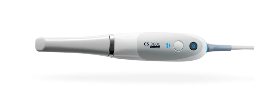
Simultaneously, invest time in researching different scanners. Explore comparisons, especially regarding accuracy and ease of handling. Reading reviews and understanding user experiences can offer valuable insights. While the decision may not be straightforward, a combination of firsthand experience and thorough research will better inform your choice.
DRA: Can you share your insights on digital implant planning and denture protocols?
IB: Implant planning is a valuable process involving the use of a CBCT scan and a scanner for creating overlays. However, it’s crucial to recognize that the accuracy of this method may not be as high as commonly perceived. Studies on implant placement with a guide reveal a certain level of failure, emphasizing the need for caution. It’s essential never to entirely rely on the accuracy of the guide. Particularly concerning intraoral scanning, there’s a need to allow for some space, as the precision may not be as absolute as assumed.
In the case of dentures, scanning, especially in the gingival areas, can be achieved effectively depending on the scanner used. However, the final step involves adding functionality to the denture, a process that, at present, I continue to execute using analog techniques.
DRA: Are there any myths about intraoral scanning or digital dentistry that you’d like to dispel?
IB: One important consideration is not to rely on advice from individuals who have only tried one scanner. Through our tests, we’ve found that some scanners are less accurate, particularly for single teeth. Therefore, it’s essential to listen to someone with experience across multiple scanners who can provide insights into the differences, not solely based on price.
While cost is a factor, the impression quality holds utmost importance, especially in daily restorative workflows. Even if a scanner is more expensive, investing in accuracy is worthwhile. Therefore, when seeking advice, it’s advisable to consult with someone who has experience with more than just one or two scanners.
DRA: Finally, you mentioned that intraoral scanning is not the future but the present. Why do you believe it’s crucial for dentists to embrace this technology now?
IB: Intraoral scanning has truly transformed me into a better dentist. Intraoral scanning and digital workflows, in my perspective, significantly enhance dentistry. This conviction is shared by colleagues who, like me, appreciate the multitude of options and capabilities it brings.
The ability to show patients visual representations, advanced diagnostics, and innovative planning that was not feasible with analog tools has elevated my practice. I firmly believe that embracing intraoral scanning is not a glimpse into the future; it is the present reality. Traditional scanning methods are, for me, already a thing of the past. It’s not just about staying current; it’s about providing the best possible dentistry to patients.
I firmly believe that embracing intraoral scanning is not a glimpse into the future; it is the present reality. Traditional scanning methods are, for me, already a thing of the past. It’s not just about staying current; it’s about providing the best possible dentistry to patients.
Dr. Ingo Baresel
I am convinced that in today’s dental landscape, not incorporating intraoral scanning limits the ability to deliver optimal treatment. While one can be a proficient dentist, the integration of techniques such as scanning images, profile assessments, caries diagnostics, and the ability to overlay situations for time comparisons and outcome simulations is paramount. Neglecting these advancements means not offering the best possible care to patients. It’s no longer a question of whether to switch or not; it’s about staying relevant and effective in the coming years.
The information and viewpoints presented in the above news piece or article do not necessarily reflect the official stance or policy of Dental Resource Asia or the DRA Journal. While we strive to ensure the accuracy of our content, Dental Resource Asia (DRA) or DRA Journal cannot guarantee the constant correctness, comprehensiveness, or timeliness of all the information contained within this website or journal.
Please be aware that all product details, product specifications, and data on this website or journal may be modified without prior notice in order to enhance reliability, functionality, design, or for other reasons.
The content contributed by our bloggers or authors represents their personal opinions and is not intended to defame or discredit any religion, ethnic group, club, organisation, company, individual, or any entity or individual.

