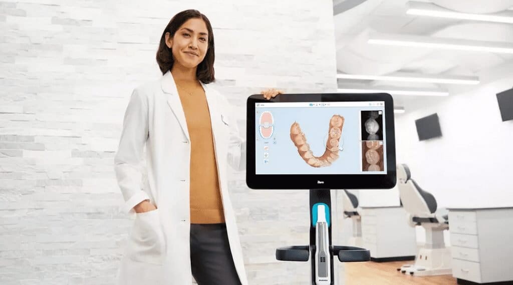
Align Technology announced the results of a multi-party clinical study that was published in the peer-reviewed Journal of Dentistry (Oct. 24, 2021), titled “Reflected near-infrared light versus bite-wing radiography for the detection of proximal caries: a multicenter prospective clinical study conducted in private practices”.
The study confirms and demonstrates the significant advantages of the iTero Element 5D imaging system in the detection and monitoring of supragingival interproximal caries without the presence of harmful radiation.

Early detection of lesions with high accuracy
The study compared near-infrared technology (NIRI) and bitewing radiographs (X-ray imaging of upper and lower molar crowns) in detecting interproximal caries. The results showed that near-infrared technology (NIRI) had high accuracy (P<0.0001) in early detection of enamel lesions (88.6%) and caries lesions at the enamel-dentin junction (96.9%).
In addition, the performance of NIRI technology and bitewing radiographs in clinically assisted removal of dental caries was compared in this study. The NIRI technology of the iTero Element 5D imaging system has shown a 66% higher sensitivity than bitewing radiographs in detecting interproximal caries lesions of posterior teeth during clinical evaluation of caries removal, and achieved 66% higher sensitivity in detecting interproximal caries in posterior teeth. 96.6% sensitivity.
“We are pleased to see the results of these clinical findings further validate what doctors and their patients have experienced.
“The visualization capabilities of the iTero Element 5D imaging system helps in the early detection of cavities without x-ray radiation,” said Yuval Shaked, senior vice president and managing director, iTero scanner and services business, Align Technology.
“This study emphasizes the valuable role that the iTero 5D imaging system with NIRI technology is already providing doctors and their staff to support their dental assessments of patients and overall patient oral healthcare and treatment options.
“When combined with the ease of use and comfortable experience for a broad population of patients, the iTero 5D imaging system with NIRI technology is an essential tool for any doctor’s office.”
NIRI technology scans the internal structure of teeth in real time
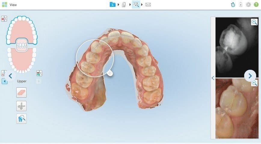
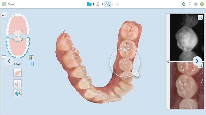
Using near-infrared technology, the internal structure of the tooth (enamel and dentin) can be scanned in real-time and a 3D color image of the tooth is created at the same time, supporting caries detection. Near-infrared images of posterior teeth were used to detect interproximal caries, and the results were compared with bitewing radiographs.

The study’s lead author, Dr. Zvi Metzger, Professor of Oral Biology and Endodontology at Tel Aviv University’s Goldschleger School of Dental Medicine, commented on the study’s results:
“Reflected near infra-red light images generated simultaneously during 3D scanning of dental arches with the iTero Element 5D imaging system scanner may be used reliably for detection, screening and monitoring of proximal caries.
“This method for caries detection may potentially minimize the traditional use of ionizing radiation.”
Said Dr. Peggy Bown, a participant in the study: “As a current user of the iTero Element 5D imaging system, participating in the study was very beneficial to me.
“I could further see the clinical value of the iTero Element 5D imaging system through its high sensitivity at detecting early enamel lesions and ease of use. Having one system, one scan, and one tip eliminates the need for multiple devices, repetitive sterilization, and minimizes the use of harmful radiation.”

Carious lesions can be detected simultaneously with every scan
Dr. Ingo Baresel, President of the German Society for Digital Intraoral Impression, who has used the imaging system in his clinic, reported: “As one of the first users of the iTero Element 5D intraoral scanner for caries diagnosis, I quickly discovered that early adjacent Facial caries are clearer than in traditional bitewing radiographs.
“This subjective impression has also been objectively confirmed by participating in this study. And because of its simplicity, I can now make better early diagnosis quickly and reliably without the need for Harmful X-rays are used. I don’t need to change the scan head to check for carious lesions in every scan.”
The information and viewpoints presented in the above news piece or article do not necessarily reflect the official stance or policy of Dental Resource Asia or the DRA Journal. While we strive to ensure the accuracy of our content, Dental Resource Asia (DRA) or DRA Journal cannot guarantee the constant correctness, comprehensiveness, or timeliness of all the information contained within this website or journal.
Please be aware that all product details, product specifications, and data on this website or journal may be modified without prior notice in order to enhance reliability, functionality, design, or for other reasons.
The content contributed by our bloggers or authors represents their personal opinions and is not intended to defame or discredit any religion, ethnic group, club, organisation, company, individual, or any entity or individual.

